Monday 17 November 2014

Lab 2: Measurement and Counting of Cells Using Microscope

2.1 Ocular Micrometer 

Introduction
An ocular micrometer is a glass disk that fits in a microscope eyepiece that has a ruled scale, which is used to measure the size of magnified objects. The physical length of the marks on the scale depends on the degree of magnification. To put it simpy, an ocular micrometer used to measure the size of object and the stage micrometer is used to calibrate the ocular micrometer. This is to ensure the accurate measurement with ocular.

Materials
Microscope fitted with an ocular micrometer
Slide micrometer
Stained preparation of yeast

Method Used
1. Place the stage micrometer on the stage.
2. Using the lowest power objective, focus the microscope until the image on the stage micrometer is observed superimposed on the eyepiece scale.
3. Determine how many divisions of the eyepiece scale correspond top a definite number of divisions
on the stage scale.
4. Calculate the measurement of an eyepiece division in micrometer (μm).
5. Calculate and record for future reference, the diameter of the field for each objective.
6. Determine the average dimensions (in μm) of a sample of yeast cells.

Results

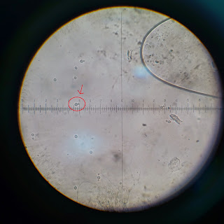
 40 magnification image seen with ocular lens

100 magnification image seen with ocular lens 

400 magnification image with yeast ( Horizontal) 

400 magnification objective lens (Vertical)

Average dimension of yeast:

10x objective lens - 1 mm = 9.5 ocular unit
40x objective lens - 0.1mm = 4 ocular unit

the length of the yeast microorganism 
4 ocular unit = 0.1 mm
0.5 ocular unit = 0.0125 mm

the width of the yeast microorganism 
4 ocular unit = 0.1 mm
0.3 ocular unit = 7.5x10^-3 mm

Discussion

-handle the yeast culture by using the aseptic technique to prepare the slide to be observed under ocular lens.
-Calibrate the ocular lens to get the measurement.
10x objective lens - 1 mm = 9.5 ocular unit
40x objective lens - 0.1mm = 4 ocular unit
-to read the width and the length,rotate the objective len.
read the micrometer carefully to avoid parallex error.

2.2 Neubauer Chamber

Introduction
The hemocytometer (or haemocytometer or counting chamber) is a specimen slide which is used to determine the concentration of cells in a liquid sample. It is frequently used to determine the concentration of blood cells (hence the name “hemo-“) but also the concentration of sperm cells in a sample. The cover glass, which is placed on the sample, does not simply float on the liquid, but is held in place at a specified height (usually 0.1mm). Additionally, a grid is etched into the glass of the hemocytometer. This grid, an arrangement of squares of different sizes, allows for an easy counting of cells. This way it is possible to determine the number of cells in a specified volume.

Materials
Serial dilutions of yeast 
Neubauer chamber and coverslip
70% ethanol 
Sterile Pasteur pipette

Method Used

1. Using a sterile Pasteur pipette, add a drop of diluted yeast culture (use 10-3 or 10-4 

2. Allow about one minute for the cells to settle dilution) to the space between the coverslip and the counting chamber. 

3. Count the cells in the four corner and centre squares. For a reasonably accurate count, you should have more than 30 cells per area.

4. Clean the Neubauer and coverslip with 70% ethanol


Counting


1. The chamber contains many grids, producing nine (9) major large squares

2. For calculation purposes, only the middle large square is used.

3. The middle large square has a size of 1 mm x 1 mm and a depth of 0.1 mm


4. Inside the middle large squares, there are 20 smaller squares, each with the size of 0.2 mm x 0.2 mm

5. Randomly choose 12 out of 20 small squares and calculate the number of yeast cells in the squares

6. Average the number of cells per square.


Results
Image of yeast on hemocytometer (40x)

total cells of 12 small boxes = 237 cells
average for 1 small box    = 237/12
                                               =19.75 cells
total cells for 400 small boxes   = 19.75 x 400
                                        = 7900
                            volume of one small box   = 1 mm x 1 mm x 0.1 mm
                                               = 0.1 mm3

1 mm3   = 0.001 cm3
0.1 mm3 = 0.0001 cm3

1 cm3  = 1 ml
0.0001 cm3  = 0.0001 ml

concentration of cells in 400 small boxes = 7900cell /0.0001 ml
                                                              = 79000000 cell/ml


Discussion


-The yeast culture is put in the hemocytometer by using aseptic techniques. The    
 coverslip must be on the hemocytometer to gives a very precise volume in the space
 delimited by the grid and the coverslip.It is being done carefully to avoid the air bubbles  
 in the counting chamber.

-The hemocytometer is thicker than a regular slide. We careful to not crash the objective
  lens into the hemocytometer at the time of focusing.

-Choose 12 boxes out of 400 boxes and calculate the number of cells in each boxes.
 Then get the average number of the cell to calculate the concentraion of the cell.
 
Conclusion 

Finally, we would like to conclude that our group members have learned the correct techniques to measure the size of yeast by using an ocular micrometer with accurate measurement. We can also count the yeast by using a hemocytometer so we can  determine the number of yeast in a specified volume.

Reference

  • http://en.wikipedia.org/wiki/Ocular_micrometer
  • http://www.wartburg.edu/biology/vlg/scale.html
  • http://en.wikipedia.org/wiki/Hemocytometer
  • http://www.microbehunter.com/the-hemocytometer-counting-chamber/


Monday 3 November 2014

Lab 1: Principle and Use of Microscope

1.1 Setting up and use of microscope

1. Introduction
"Micro" refers to tiny, "scope" refers to view or look at. Microscopes are tools used to enlarge images of small objects so as they can be studied. Microscopes range from a simple magnifying glass to the expensive electron microscope. The compound light microscope is the most common instrument used. It is an instrument containing two lenses, which magnifies, and a variety of knobs to resolve (focus) the picture. It is a rather simple piece of equipment to understand and use. 

2. Materials

A microscope 
Glass slide
Cover slip
3. Method Used 

Setting up a microscope
1. Sit on your stool with your knee under the bench and move the microscope so that you look through 
both eyepieces without straining. Make sure you are comfortable.
2. After plugging in the power lead of the microscope and turning on the power, turn on the microscope 
light using the main on-off switch.
3. Adjust the light intensity using the brightness control. Position 5 is normally adequate.
4. Rotate the revolving nosepiece to bring the 4x objective lens into the light path.
5. Take a clean slide and mark a line on it with a marker pen. Place the slide on the stage, using the 
spring clip to secure it. Move the slide into the light path using the coaxial stage control knobs.
6. Look through both eyepieces and adjust them until you see a single circle of light. For future 
reference, make a note in your class manual of the setting on the interpupillary distance scale.
7. Rotate the tube length adjustment (diopter) ring on the right eyepiece to match your interpupillary 
distance setting obtained in 1.6.
8. Using the right eye only, focus the marker-pen mark by adjusting the coarse and fine adjustment 
knobs.
9. Using the left eye only, focus the left eyepiece using the tube lens adjustment (diopter) ring. Again 
for future, make a note in class manual of the diopter ring setting

1.2 Examination of Cells

1. Introduction

Some samples can be placed directly under the microscope. However, many samples look better when placed in a drop of water on the microscope slide. This is known as a "wet mount." The water helps support the sample and it fills the space between the cover slip and the slide allowing light to pass easily through the slide, the sample, and the cover slip. 

2. Materials
Culture
Immersion oil
Lens tissue
A microscope slide containing stained microorganisms
Inoculating loop
Bunsen burner
Slide and coverslip
3. Procedure
Stained cells:

1. Set up microscope as described above and examine the slide under the oil immersion lens.
2. Observe the shape and size of the organisms and any structure that are visible. Draw what you see.
The wet mount:

1. Use a sterile Pasteur pipette to aseptically transfer one drop of culture to the centre of a glass slide.
2. Use a marker pen to mark a coverslip. This mark will help you to focus on the microorganisms.
3. Take the coverslip and turn it so that the marker pen mark faces down. Then place one edge of the 
coverslip onto the slide and gently lower it so that it covers the drop of culture. The culture will 
spread between the coverslip and the slide.
4. Place the slide on the microscope stage and using the 4x objective focus on the culture. You may see 
two or three groups of highly motile protozoa.
5. Observe the cell using the 10x and the 40x objectives. By observing closely you may be able to 
detect some smaller moving objects. They will be some of the larger types of bacteria. Make drawing 
of what you see.
6. Observe the cells using oil immersion lens. Remember to adjust the condenser and diaphragm. Again 
make drawings and comment on anything of interest you see.
7. Repeat this procedure with other cultures. 
4. Results
Stained Cell under Microscope

1) Bacteria type: Cocci, Bacilli, Spirilli 

 (40x magnification)

100x magnification


400x magnification

Oil immersion, 1000x magnification

2) Typical Bacillus 

40x magnification 

100x magnification  

400x magnification 

 Oil Immersion 1000x magnification

3) Penicillium Conidia


40x magnification 

100x magnication  
400x magnification

1000x magnification 


Wet Mount:
Lactobacillus fermentum
 

                                                              1000x  magnification


Discussion: 

The morphology of Lactobacillus fermentum :
-Shape: Rod
-Size: Tiny
-Surface: Smooth
-Texture: Moist

The morphology of Typical Bacillus
-Shape: Rod
-Size: Tiny
-Surface: Smooth
-Texture: Moist

The morphology of Penicillium Conidia 
-Shape: highly branched network of multinucleate, septate, usually colorless hyphae
-Size: Medium
-Surface: Smooth

Conclusion: 

As a conclusion, i have learned the right techniques to use a microscope. I also learned the right way to practice aseptic technique to avoid contamination to occur while handling cultures. I have obtained a clear view on the morphology of the microorganisms like Lactobacillus fermentum in this experiment by using immersion oil which could ensure magnification of 1000x achieved while still preserving good resolution.

Reference: 
  • http://en.wikipedia.org/wiki/Bacillus_(shape)
  • http://en.wikipedia.org/wiki/Penicillium
  • http://en.wikipedia.org/wiki/Microscope
  • http://ibg102.wordpress.com/2013/04/05/lab-1-principles-and-use-of-microscope/